New possibilities of exploring artworks with X-ray transmission radiography using the latest generation of pixel detectors
InsightART, U Pergamenky 12, Prague 7, 170 00
Abstract
X-ray transmission radiography is one of the well-known and commonly used non-destructive methods in the exploration of works of art. X-ray radiation is used by restorers, conservators, technologists, and other professionals in the industry. However, the latest generation of particle detectors, which InsightART uses to analyze works of art, opens up entirely new possibilities in the field of X-ray radiography and its post-production.
Currently, photographic films, or CCD or CMOS cameras with scintillators, are still used for X-ray imaging. However, these devices only measure the intensity of radiation. In addition, these technologies suffer from internal noise that limits the maximum dynamic range (number of greyscale levels), thus reducing the resolution of tiny details in the image. The new generation of pixel detectors significantly advances the possibilities of X-ray imaging.
The state-of-the-art pixel detectors used by InsightART are equipped with so-called smart pixels. Each of these pixels contains electronics to measure the wavelength of each detected photon. The latest “RToo” scanners thus produce completely unprecedented outputs in the analysis of artworks. The resulting X-rays are fully digital, colour, and high resolution (over 100 MPix). However, the greatest benefit of this method is the ability to distinguish individual materials in the analyzed works. The new scanner, developed in collaboration with Radalytica s.r.o., uses robotic arms to move, allowing unique flexibility in scanning 3D objects such as sculptures, furniture, or archaeological finds.
Introduction
The properties of the new generation of X-ray imaging detectors allow significant improvements in the quality of X-rays in terms of resolution and contrast. The principle of obtaining X-ray images remains the same (Fig. 1). The difference is the imaging device, which offers new properties.

The most important extension of X-ray imaging capabilities is the ability of these detectors to analyze only the selected wavelength of radiation. These are, therefore, hyperspectral pixel detectors, as already used in the visible spectrum, with the difference that they operate in the X-ray spectrum. Measurement of the X-ray spectrum variation passing through the object under investigation offers the possibility of recognizing the individual materials from which the object is composed.
These properties provide an insight into the image in its microscopic dimensions, allowing very accurate mapping of the object under investigation.
In spectral (i.e. energy-sensitive) transmission radiography, we can assign colours to individual differentiated materials so that they can be distinguished in the resulting X-ray image. Different colours thus represent a material or a group of similar materials (pigments). The result is a high-resolution X-ray material map that gives invaluable information about its internal structure.
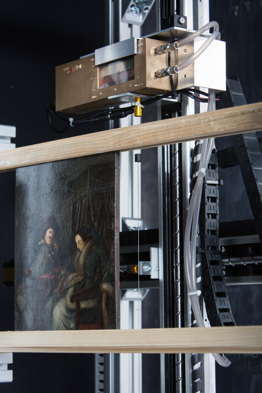
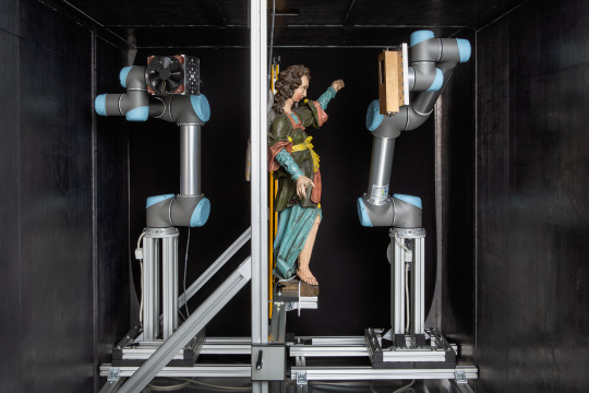
Currently, InsightART has RToo 2D (Fig. 2) and RToo 3D (Fig. 3) scanning systems. Both devices use Widepix 1×5 detectors manufactured by the Czech company ADVACAM, s.r.o. These detectors are based on Medipix/Timepix technology, developed in a broad international collaboration led by CERN in Geneva. Widepix detectors have an array of 256 x 1280 pixels at 55 μm. Because they are single-photon imaging detectors, the measured data is not loaded with electronic noise, allowing practically any contrast and dynamic range.
The 2D model is primarily designed for scanning paintings, i.e. paintings on canvas, wooden boards, etc.
The 3D model already uses a system of two robot manipulators to allow continuous synchronized movement in 3D space, opening up the possibility of scanning 3D artwork, such as polychromed wood sculptures, ceramic artefacts, etc.
X-ray transmission radiography in high resolution
In order to fully appreciate the benefits of RTG radiographic methods in the field of scanning artwork using spectral pixel detectors, it is necessary to understand the scope of practical application. As with healthcare, where it is necessary to identify the extent and type of illness as accurately as possible in order to establish adequate treatment, in the restoration and authentication of works of art we can also apply accurate and correct processes only if the initial investigation is consistent. Comparison to the medical environment is appropriate as both disciplines use very similar methods of examination – both physical and chemical. However, the highest degree of concordance lies in the elimination of invasive procedures of applied analysis and the need to improve and refine the achievable results of applied detection methods. In other words, the results of examining the physical structure of matter are only as good and accurate as the apparatus we are examining it with.
The more accurate and detailed our knowledge, the better we can determine the origin or age of the object under investigation. The use of spectral X-ray pixel detectors in the medical and artistic-historical fields is a great step forward from this perspective.
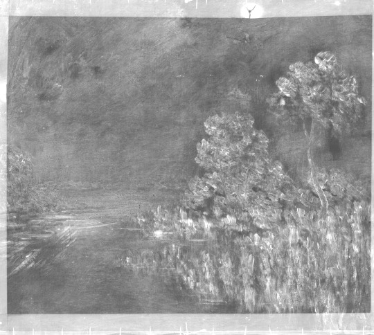
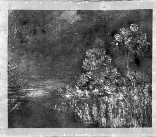
When comparing black-and-white X-rays using photographic film and the Widepix detector, the distinctly higher image quality in contrast and dynamic range is immediately apparent (Fig. 4). In the post-production process and subsequent radiogram analysis, a high-resolution image is preferred (Fig. 5).
Material-sensitive X-ray radiography and its benefits
For non-invasive exploration of paintings, this method is a very significant asset in terms of a broad range of usable information. Full surface material radiographic mapping of the work without the need for invasive intervention in its structure is therefore the most significant benefit of Widepix spectral pixel detectors. This method of full surface material mapping brings entirely new possibilities to the field of artwork exploration. Firstly, it gives the researcher perfect orientation in the internal structure of the work due to the high resolution of the measured radiographic image. It also provides knowledge of the distribution of individual materials throughout the whole image area and in all its layers, again in high resolution, which allows the work to be examined in microscopic dimensions.
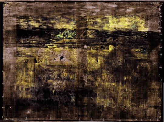
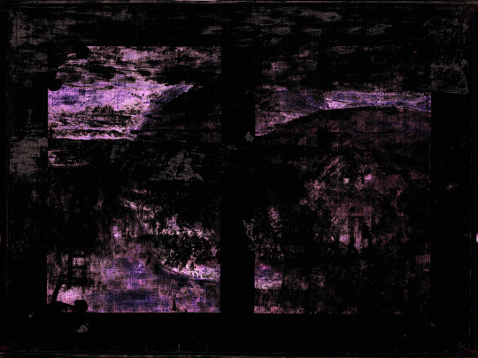
This method also has great potential in the possibility of postproduction images of just certain measured types of materials that relate to individual pigments. In this way, we can see, for example, only the presence of lead pigments on the whole surface of the image, or use a combination of pigments on the basis of iron, titanium, copper, etc. Using material-sensitive X-ray radiography in this way opens up entirely new possibilities not only for precise setting of restoration and preservation processes, but primarily for the authentication of works of art and for the detection of fakes (Fig. 6 to Fig. 8).
Selecting certain colour channels (materials) in the resulting radiographic image can also serve to uncover different author changes and adjustments in the composition of the image. For many artists, it is also common to repaint their own paintings with other motifs; in such cases it is possible to visually “separate” individual layers from one another by means of spectral information.
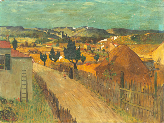
La Crau with a View of Montmajour
The painting of La Crau with a View of Montmajour (Fig. 9) is a unique example of the use of material-sensitive X-ray radiography using RToo imaging equipment with Widepix detector. It is an oil painting on canvas, showing a view of the landscape near the French Arles with the ruins of Montmajour Abbey in the background. In the lower left corner is the signature Vincent. The image was purchased by the current owner in an auction, where it was listed as a copy by van Gogh. With its visual style and thematic focus this work fits the style of Vincent van Gogh’s work in 1888.
However, there is only a minimal amount of information about the origin of this work and, in the past, there has never been a thorough restoration or technological and historical research into the painting. For these reasons, in 2015-2018 the work was subject to a full range of available analyses including material-sensitive X-ray radiography.
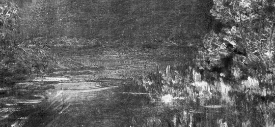
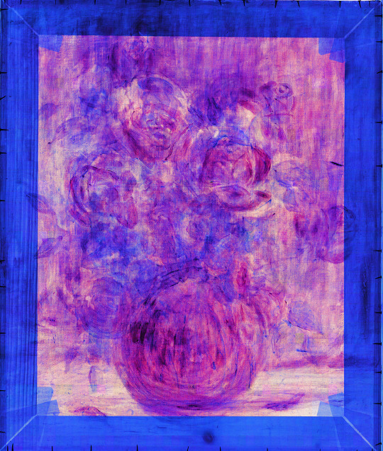
During the radiographic survey of the work, it was found that there is another image under the painting of the La Crau landscape, namely the figure of a female nude shown from behind (Fig. 11). This figure was commenced only with brown tones in the shadow parts and white pastes in the illuminated parts of the figure. The style of underpainting and the position of the model show that it is an incomplete study painting of the nude, similar to the models van Gogh drew and painted in classes of the local academy in 1886-88, during his Parisian visit. The paste layers of the underpainting were partially scraped before painting the new image, and thus on the surface of the current painting a small relief is visible.
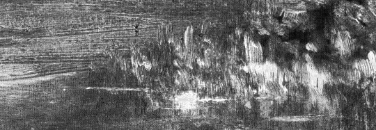
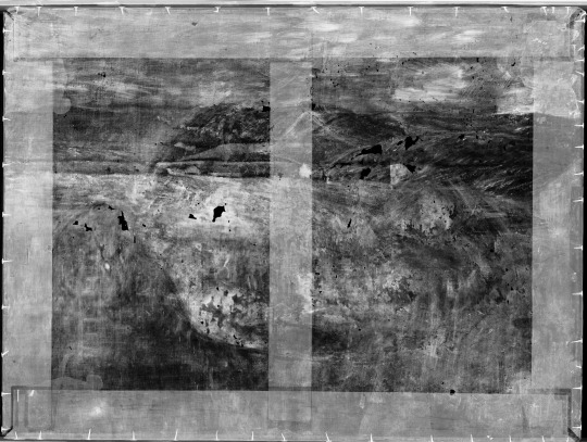
The material-sensitive X-ray images show the exact distribution of individual pigments across the image area, as well as the way they are applied in individual parts of the work. Using the separation of individual energy channels, we can get an accurate survey of the author’s work with individual layers and materials (Fig. 12 and Fig. 13). In the subsequent post-production process, it is also possible to show materials that are only related to the layer of paint that we want to visualize (Fig. 14).
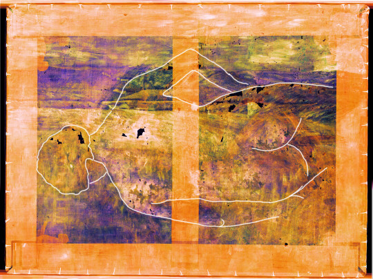
Only the original figure of the female nude is shown in the picture; all the paint layers from the top landscape painting of La Crau with a View of Montmajour were suppressed here by filtering the signal in the individual energy channels. Thus, it is evident that this method allows a completely new way of working with X-ray radiography using the most advanced pixel detectors. The post-production image processing options are enormous and bring with them many ways to process the resulting X-ray image so that they have the greatest possible value for the studied work. For example, the exact representation of a figure in lower layer of La Crau with a View of Montmajour allows a precise comparison to be made of the presumed author’s painting style with his/her other works (Fig. 15 and Fig. 16).
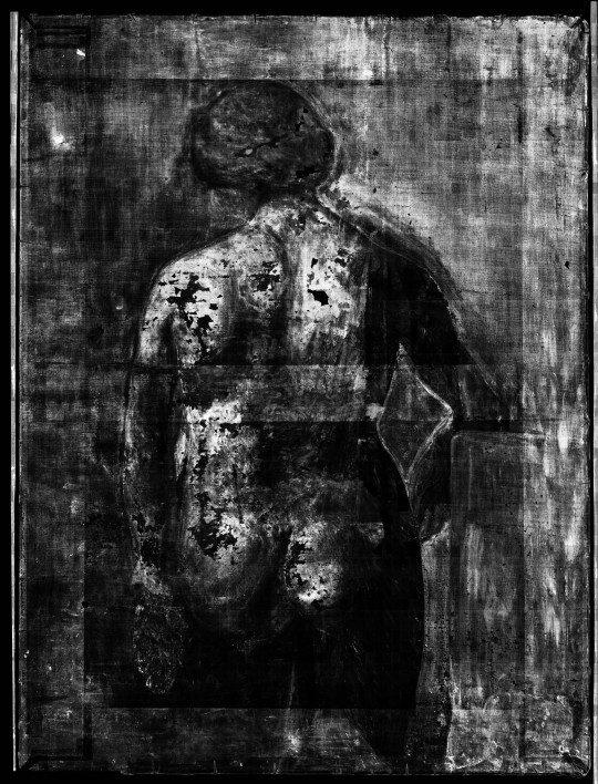


Fig. 15 a) b) c)
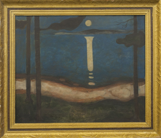
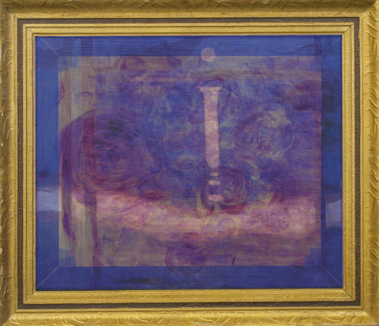
Van Gogh’s studies of a female nude of 1886-7.

Conclusion
It is clear that X-ray transmission radiography using the latest pixel detectors has a great future in the field of artwork research. By engaging other technological processes, such as artificial intelligence for precise determination of detected pigments in paints, or virtual reality for more thorough analysis of radiographs, this method is becoming an important tool for accurate identification of artworks in the authentication process. In terms of extensive application and utilization capabilities, it is evident that these methods will soon appear in the everyday practice of restorers, conservators, technologists and historians, who will significantly benefit from them in their profession. Obviously, the development of these technologies continues; however, at present we already have a new functional tool that significantly shifts the field of X-ray radiography.